Studies at the Intersection of Ribosome Function and Cellular Homeostasis

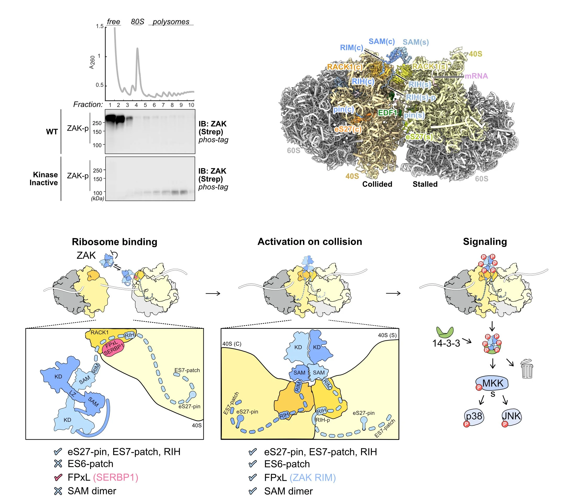
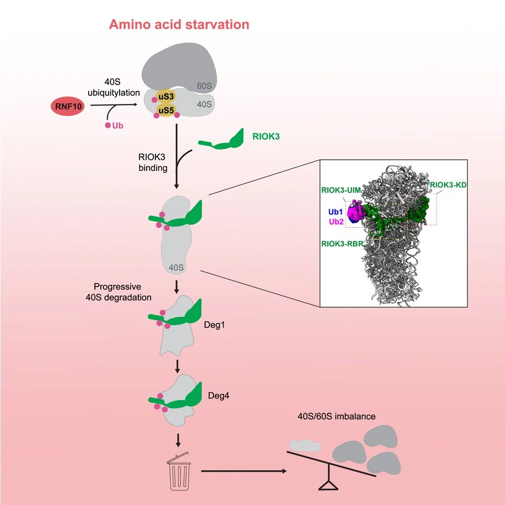
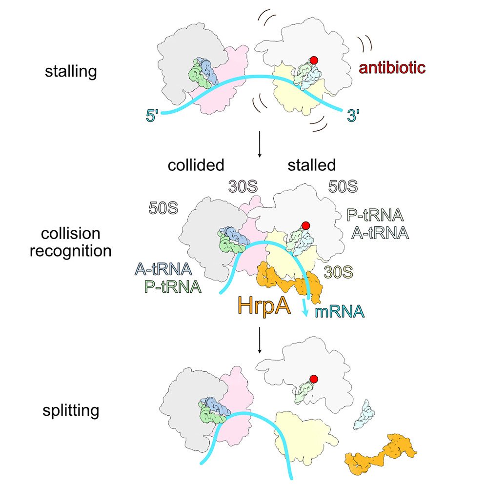
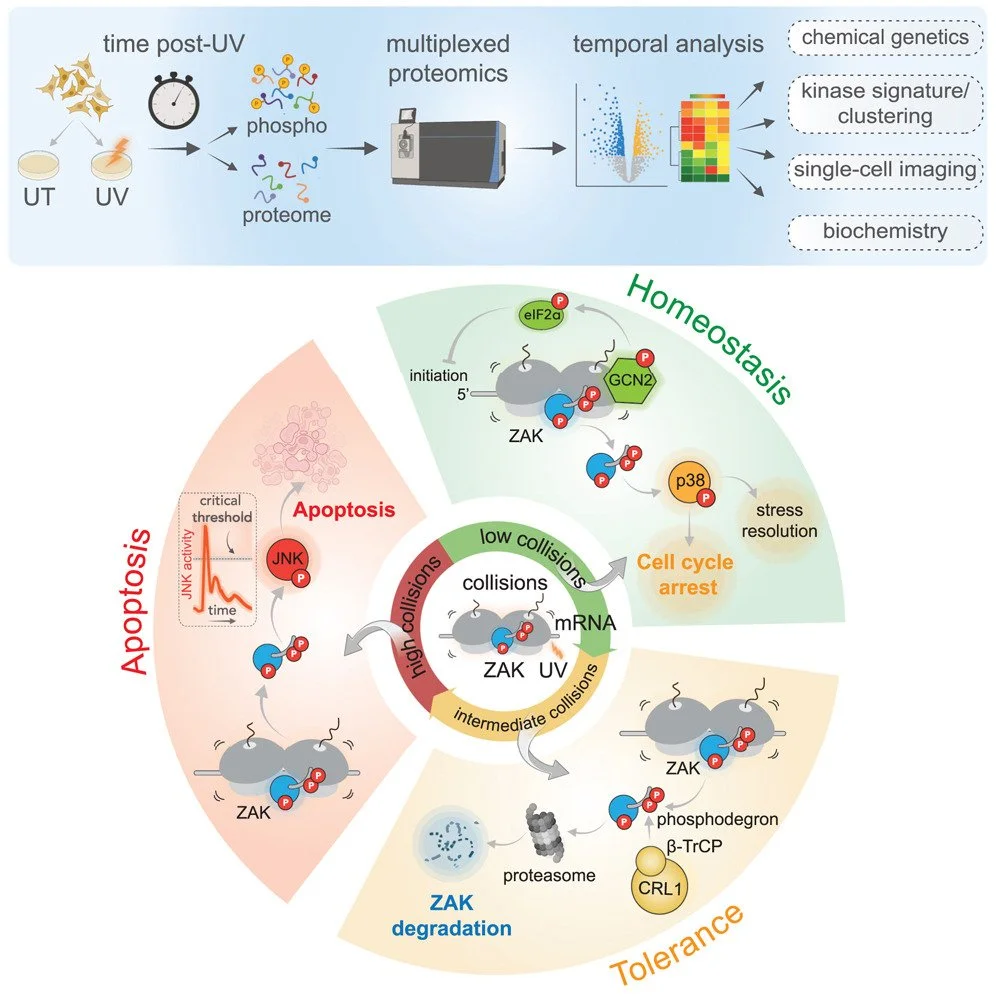
We are broadly interested in the role of ribosomes in cells in translating mRNAs into functional polypeptides and in the regulation of translation in responding to signals from the outside world. Our most recent work has focused on ribosome-mediated quality control and the many factors implicated in this process. We have used genetic and proteomic screens in yeast, mammals and bacteria to identify novel factors involved in these pathways. Once we identify factors of interest, we use a broad range of approaches to characterize the role of these factors in the process with a strong emphasis on molecular mechanism. We have shown that many of these factors specifically recognize collided ribosomes, thus revealing a unifying principle for how cells must assess overall translational distress. We routinely collaborate with cryoEM labs to leverage structural information in our studies and recently published the first atomic resolution structure of a quality control factor (EDF1/Mbf1) bound to colliding ribosomes.

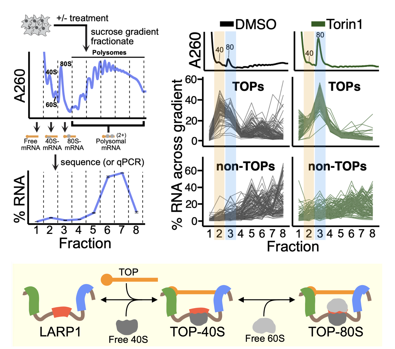
We recently have identified direct connections between ribosome collisions and cellular signaling pathways and we are exploring these connections at the molecular level. Ongoing work in this area is defining the interactions of ribosome-mediated quality control and cellular signaling pathways in tissues where protein synthesis regimes may be different (skin, cancer cells, neurons). Our interests in ribosome recycling and rescue have raised our interest in ribosome homeostasis and how it contributes to the regulation of gene expression. Why do ribosome deficiencies in different tissues lead to differential sensitivities to these deficiencies? How does the cell balance the demands of protein synthesis with the regulation of initiation, elongation, termination, and recycling steps of this process to ensure that gene expression happens appropriately in order to sustain the life of the organism? We also continue to be interested in connections between ribosome read-through at stop codons and nonsense-mediated decay, based in part on the relevance of such models to therapeutic treatment for diseases originating from premature stop codons in critical genes.

ZAK activation at the collided ribosome

Ribosome collisions activate the ribotoxic stress response mediated by the MAP3K ZAK, which in turn regulates cell-fate consequences through downstream phosphorylation of the MAPKs p38 and JNK. Despite the critical role of ZAK during cellular stress, a mechanistic and structural understanding of ZAK–ribosome interactions and how these lead to activation remain elusive. Here we combine biochemistry and cryo-electron microscopy to discover distinct ZAK–ribosome interactions required for constitutive recruitment and for activation. We find that upon induction of ribosome collisions, interactions between ZAK and the ribosomal protein RACK1 enable its activation by dimerization of its SAM domains at the collision interface. Furthermore, we discover how this process is negatively regulated by the ribosome-binding protein SERBP1 to prevent constitutive ZAK activation. Characterization of novel SAM variants as well as a known pathogenic variant of the SAM domain of ZAK supports a key role of the SAM domain in regulating kinase activity on and off the ribosome, with some mutants bypassing the ribosome requirement for ZAK activation. Collectively, our data provide a mechanistic blueprint of the kinase activity of ZAK at the collided ribosome interface. (Huso* Niu* et al, Nature, 2025)


RIOK3 mediates the degradation of 40S ribosomes

Cells tightly regulate ribosome homeostasis to adapt to changing environments. Ribosomes are degraded during stress, but the mechanisms responsible remain unclear. Here, we show that starvation induces the selective depletion of 40S ribosomes following their ubiquitylation by the E3 ligase RNF10. The atypical kinase RIOK3 specifically recognizes these ubiquitylated 40S ribosomes through a unique ubiquitin-interacting motif, visualized by cryoelectron microscopy (cryo-EM). RIOK3 binding and ubiquitin recognition are essential for 40S ribosome degradation during starvation. RIOK3 induces the degradation of ubiquitylated 40S ribosomes through progressive decay of their 18S rRNA beginning at the 3′ end, as revealed by cryo-EM structures of degradation intermediates. Together, these data define a pathway and mechanism for stress-induced degradation of 40S ribosomes, directly connecting ubiquitylation to regulation of ribosome homeostasis. (Huang* Diehl* et al, Molecular Cell, 2025)


The RNA helicase HrpA rescues collided ribosomes in E. coli

Although many antibiotics inhibit bacterial ribosomes, the loss of known factors that rescue stalled ribosomes does not lead to robust antibiotic sensitivity in E. coli, suggesting the existence of additional mechanisms. Here, we show that the RNA helicase HrpA rescues stalled ribosomes in E. coli. Acting selectively on ribosomes that have collided, HrpA uses ATP hydrolysis to split stalled ribosomes into subunits. Cryoelectron microscopy (cryo-EM) structures reveal how HrpA simultaneously binds to two collided ribosomes, explaining its selectivity, and how its helicase module engages downstream mRNA such that, by exerting a pulling force on the mRNA, it would destabilize the stalled ribosome. These studies show that ribosome splitting is a conserved mechanism that allows proteobacteria to tolerate ribosome-targeting antibiotics. (Campbell et al, Molecular Cell, 2025)


LARP1 binds ribosomes and TOP mRNAs in repressed complexes

Terminal oligopyrimidine motif-containing mRNAs (TOPs) encode all ribosomal proteins in mammals and are regulated to tune ribosome synthesis to cell state. Previous studies have implicated LARP1 in 40S- or 80S-ribosome complexes that are thought to repress and stabilize TOPs. However, a molecular understanding of how LARP1 and TOPs interact with these ribosome complexes is lacking. Here, we show that LARP1 directly binds non-translating ribosomal subunits. Cryo-EM structures reveal a previously uncharacterized domain of LARP1 bound to and occluding the mRNA channel of the 40S subunit. Increased availability of free ribosomal subunits downstream of various stresses promote 60S joining at the same interface to form LARP1-80S complexes. Simultaneously, LARP1 engages the TOP via its previously characterized La/PAM2 and DM15 domains. Contrary to expectations, ribosome binding within these complexes is not required for LARP1-mediated TOP repression or stabilization, two canonical LARP1 functions. Together, this work provides molecular insight into how LARP1 directly binds ribosomal subunits and challenges existing models describing the function of repressed LARP1-40S/80S-TOP complexes. (Saba et al, EMBO, 2024)


The ribotoxic stress response drives UV-mediated cell death

While ultraviolet (UV) radiation damages DNA, eliciting the DNA damage response (DDR), it also damages RNA, triggering transcriptome-wide ribosomal collisions and eliciting a ribotoxic stress response (RSR). However, the relative contributions, timing, and regulation of these pathways in determining cell fate is unclear. Here we use time-resolved phosphoproteomic, chemical-genetic, single-cell imaging, and biochemical approaches to create a chronological atlas of signaling events activated in cells responding to UV damage. We discover that UV-induced apoptosis is mediated by the RSR kinase ZAK and not through the DDR. We identify two negative-feedback modules that regulate ZAK-mediated apoptosis: (1) GCN2 activation limits ribosomal collisions and attenuates ZAK-mediated RSR and (2) ZAK activity leads to phosphodegron autophosphorylation and its subsequent degradation. These events tune ZAK's activity to collision levels to establish regimes of homeostasis, tolerance, and death, revealing its key role as the cellular sentinel for nucleic acid damage. (Sinha et al, Cell, 2024)


EDF1 coordinates cellular responses to ribosome collisions


Translation of aberrant mRNAs induces ribosomal collisions, thereby triggering pathways for mRNA and nascent peptide degradation and ribosomal rescue. Here we use sucrose gradient fractionation combined with quantitative proteomics to systematically identify proteins associated with collided ribosomes. This approach identified Endothelial differentiation-related factor 1 (EDF1) as a novel protein recruited to collided ribosomes during translational distress. Cryo-electron microscopic analyses of EDF1 and its yeast homolog Mbf1 revealed a conserved 40S ribosomal subunit binding site at the mRNA entry channel near the collision interface. EDF1 recruits the translational repressors GIGYF2 and EIF4E2 to collided ribosomes to initiate a negative-feedback loop that prevents new ribosomes from translating defective mRNAs. Further, EDF1 regulates an immediate-early transcriptional response to ribosomal collisions. Our results uncover mechanisms through which EDF1 coordinates multiple responses of the ribosome-mediated quality control pathway and provide novel insights into the intersection of ribosome-mediated quality control with global transcriptional regulation. (Sinha et al, Elife, 2020)

Ribosome collisions trigger general stress responses to regulate cell fate

Problems arising during translation of mRNAs lead to ribosome stalling and collisions that trigger a series of quality control events. However, the global cellular response to ribosome collisions has not been explored. Here, we uncover a function for ribosome collisions in signal transduction. Using translation elongation inhibitors and general cellular stress conditions, including amino acid starvation and UV irradiation, we show that ribosome collisions activate the stress-activated protein kinase (SAPK) and GCN2-mediated stress response pathways. We show that the MAPKKK ZAK functions as the sentinel for ribosome collisions and is required for immediate early activation of both SAPK (p38/JNK) and GCN2 signaling pathways. Selective ribosome profiling and biochemistry demonstrate that although ZAK generally associates with elongating ribosomes on polysomal mRNAs, it specifically auto-phosphorylates on the minimal unit of colliding ribosomes, the disome. Together, these results provide molecular insights into how perturbation of translational homeostasis regulates cell fate. (Wu et al, Cell, 2020)

Ribosome collisions induce mRNA cleavage and ribosome rescue in bacteria


Ribosome rescue pathways recycle stalled ribosomes and target problematic mRNAs and aborted proteins for degradation. In bacteria, it remains unclear how rescue pathways distinguish ribosomes stalled in the middle of a transcript from actively translating ribosomes. Here, using a genetic screen in Escherichia coli, we discovered a new rescue factor that has endonuclease activity. SmrB cleaves mRNAs upstream of stalled ribosomes, allowing the ribosome rescue factor tmRNA (which acts on truncated mRNAs) to rescue upstream ribosomes. SmrB is recruited to ribosomes and is activated by collisions. Cryo-electron microscopy structures of collided disomes from E. coli and Bacillus subtilis show distinct and conserved arrangements of individual ribosomes and the composite SmrB-binding site. These findings reveal the underlying mechanisms by which ribosome collisions trigger ribosome rescue in bacteria.

Translational repression of NMD targets by GIGYF2 and EIF4E2

Translation of messenger RNAs (mRNAs) with premature termination codons produces truncated proteins with potentially deleterious effects. This is prevented by nonsense-mediated mRNA decay (NMD) of these mRNAs. NMD is triggered by ribosomes terminating upstream of a splice site marked by an exon-junction complex (EJC), but also acts on many mRNAs lacking a splice junction after their termination codon. We developed a genome-wide CRISPR flow cytometry screen to identify regulators of mRNAs with premature termination codons in K562 cells. This screen recovered essentially all core NMD factors and suggested a role for EJC factors in degradation of PTCs without downstream splicing. Among the strongest hits were the translational repressors GIGYF2 and EIF4E2. GIGYF2 and EIF4E2 mediate translational repression but not mRNA decay of a subset of NMD targets and interact with NMD factors genetically and physically. Our results suggest a model wherein recognition of a stop codon as premature can lead to its translational repression through GIGYF2 and EIF4E2.

Translational initiation in E. coli occurs at the correct sites genome-wide in the absence of mRNA-rRNA base-pairing 


Shine-Dalgarno (SD) motifs are thought to play an important role in translational initiation in bacteria. Paradoxically, ribosome profiling studies in E. coli show no correlation between the strength of an mRNA’s SD motif and how efficiently it is translated. Performing profiling on ribosomes with altered anti-Shine-Dalgarno sequences, we find that the mutant ribosomes select start sites correctly, arguing that start sites are hard-wired for initiation through the action of other mRNA features. Our genome-wide study reveals that SD motifs are not necessary for ribosomes to determine where initiation occurs, though they do affect how efficiently initiation occurs. (Saito et al, Elife, 2020)

The endonuclease Cue2 cleaves mRNAs at stalled ribosomes during No Go Decay


Translation of problematic sequences in mRNAs leads to ribosome collisions that trigger a series of quality control events including ribosome rescue, degradation of the stalled nascent polypeptide, and targeting of the mRNA for decay (No Go Decay or NGD). Using a reverse genetic screen in yeast, we identify Cue2 as the conserved endonuclease that is recruited to stalled ribosomes to promote NGD. Ribosome profiling and biochemistry provide strong evidence that Cue2 cleaves mRNA within the A site of the colliding ribosome. We demonstrate that NGD primarily proceeds via Xrn1-mediated exonucleolytic decay and Cue2-mediated endonucleolytic decay normally constitutes a secondary decay pathway. Finally, we show that the Cue2-dependent pathway becomes a major contributor to NGD in cells depleted of factors required for the resolution of stalled ribosome complexes. Together these results provide insights into how multiple decay processes converge to process problematic mRNAs in eukaryotic cells.​ (D’Orazio et al, Elife, 2019)

Live-cell imaging reveals kinetic determinants of quality control triggered by ribosome stalling

Translation of problematic mRNA sequences induces ribosome stalling, triggering quality-control events, including ribosome rescue and nascent polypeptide degradation. To define the timing and regulation of these processes, we developed a SunTag-based reporter to monitor translation of a problematic sequence (poly[A]) in real time on single mRNAs. Although poly(A)-containing mRNAs undergo continuous translation over the timescale of minutes to hours, ribosome load is increased by ∼3-fold, reflecting long queues of ribosomes extending far upstream of the stall. We monitor the resolution of these queues in real time and find that ribosome rescue is very slow compared to both elongation and termination. Modulation of pause strength, collision frequency, and the collision sensor ZNF598 reveals how the dynamics of ribosome collisions and their recognition facilitate selective targeting for quality control. Our results establish that slow clearance of stalled ribosomes allows cells to distinguish between transient and deleterious stalls. (Goldman et al, Mol Cell, 2021)

High-resolution ribosome profiling defines discrete ribosome elongation states and translational regulation during cellular stress

Ribosomes undergo substantial conformational changes during translation elongation to accommodate incoming aminoacyl-tRNAs and translocate along the mRNA template. We used multiple elongation inhibitors and chemical probing to define ribosome conformational states corresponding to differently sized ribosome-protected mRNA fragments (RPFs) generated by ribosome profiling. We show, using various genetic and environmental perturbations, that short 20–22 or classical 27–29 nucleotide RPFs correspond to ribosomes with open or occupied ribosomal A sites, respectively. These distinct states of translation elongation are readily discerned by ribosome profiling in all eukaryotes we tested, including fungi, worms, and mammals. This high-resolution ribosome profiling approach reveals mechanisms of translation-elongation arrest during distinct stress conditions. Hyperosmotic stress inhibits translocation through Rck2-dependent eEF2 phosphorylation, whereas oxidative stress traps ribosomes in a pre-translocation state, independent of Rck2-driven eEF2 phosphorylation. These results provide insights and approaches for defining the molecular events that impact translation elongation throughout biology. (Wu et al, Mol Cell, 2019)

Stop codon context influences genome-wide stimulation of termination codon readthrough by aminoglycosides

Stop codon readthrough (SCR) occurs when the ribosome miscodes at a stop codon. Such readthrough events can be therapeutically desirable when a premature termination codon (PTC) is found in a critical gene. To study SCR in vivo in a genome-wide manner, we treated mammalian cells with aminoglycosides and performed ribosome profiling. We find that in addition to stimulating readthrough of PTCs, aminoglycosides stimulate readthrough of normal termination codons (NTCs) genome-wide. Stop codon identity, the nucleotide following the stop codon, and the surrounding mRNA sequence context all influence the likelihood of SCR. In comparison to NTCs, downstream stop codons in 3′UTRs are recognized less efficiently by ribosomes, suggesting that targeting of critical stop codons for readthrough may be achievable without general disruption of translation termination. Finally, we find that G418-induced miscoding alters gene expression with substantial effects on translation of histone genes, selenoprotein genes, and S-adenosylmethionine decarboxylase (AMD1). (Wangen et al, Elife, 2020)